How Optical Microscopy Solved a Common Lab Problem
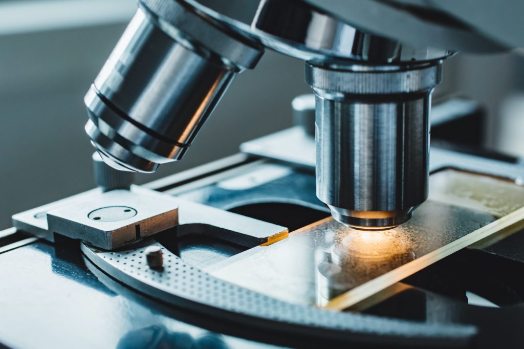
Researchers routinely study the behaviour of biological molecules – particularly proteins – beneath a microscope, in the hopes of identifying a particular protein’s function. In an effort to make tracing a protein easier, researchers often add a molecular label. Far from the traditional paper labels, most people imagine when presented with the term, molecular labels utilize a dye or covalent bonds to attach a fluorescent, radioactive, or reporter enzyme molecule to the target protein.
Understanding the pros and cons of labelled imaging can reveal why methods like microscopy are excellent for protein characterization and tracking protein movement within a live cell.
Drawbacks of Labeled Imaging
While such markers make following a target protein much easier for researchers, there are drawbacks as well. Some dyes and fluorescents interfere with regular cell function, which may make the study of protein behaviour irrelevant. Others provide too much fluorescence, making it difficult to distinguish between the protein and other cell parts, or can even be subject to photobleaching from surrounding light.
As a result, researchers have developed ways to study tissue structure, protein behaviour, and other phenomena without the need to add fluorescent labels or dyes to lab samples. These label-free imaging techniques eliminate the negative effects more intrusive labelling has on the sample itself as well as on the imaging generated. Primarily, these techniques rely on the use of optical microscopy.
Optical Microscopy
The use of optical microscopy to study cell and protein behaviour is not new. However, advances in the lasers used to produce images of the material studied has led to a drastic increase in the quality of the images provided. In addition, the imaging is non-invasive and produces very little effect on the cell materials studied.
Primarily, these achievements necessitated the use of two-photon excited fluorescence microscopy, a technology that uses a precise, ultrafast laser to provide a contrast between proteins or other cell parts. This laser usually operates at an infrared level, pulsing two or more photons very quickly to excite fluorophore compounds. In response, these compounds release a photon, emitting light on the visible spectrum.
Choosing the Right Technique
As with any light-emitting procedure, scatter – the re-absorption and re-emitting of the light produced by other, surrounding compounds – is bound to occur. Some label-free methods, such as optical coherence tomography, utilize this scatter to reconstruct the image produced from the scatter pattern. Other methods, like second harmonic generation microscopy (SHG) and third harmonic generation microscopy (THG), involve a nonlinear light scattering process, while infrared microscopy causes the very chemical bonds in a molecule to vibrate.
Another exciting technique is interferometric scattering microscopy, which works by collecting the scattered light together with a reference field of light. Researchers measure the interference caused by the light scatter with the reference provided by a laser, allowing imaging of proteins as small as 50 kilodaltons.
Depending on the research target, one technique may be preferable to another. For example, THG is fairly versatile but cannot be used extensively in live samples because its ionizing radiation can irreversibly damage cells. It is important as well for labs to choose the correct wavelength for the work at hand since certain wavelengths minimize photodamage and are preferable for time-lapse imaging.
Continued Progress
Many labs combine the use of two, three, or more of these techniques, depending on the results they are seeking to achieve. As research and development within the optical microscopy field continues, scientists hope to refine multimodal techniques to give labs the option to utilize all the best aspects of each technique, together, and without interference from one another. Continued progress in these efforts could lead to a minimally invasive, non-interfering, label-free way to gain the most information possible from a given sample.
Sources:
https://www.nature.com/articles/s41592-019-0664-8
https://en.wikipedia.org/wiki/Interferometric_scattering_microscopy
https://www.toptica.com/applications/optical-microscopy/multiphoton-microscopy/


Responses