Observing Microorganism (B)

Part 2 – STAINING

Microorganisms are observed and studied with the help of microscopes. However the bacteria have nearly the same refractive index as water, which makes them visually opaque. Hence, the microbial cells are routinely stained for clear microscopic examination. Staining is thus an auxiliary technique used in microscopy and microbiology to enhance contrast in the microscopic images of the microorganisms. Different types of stains and staining procedures are available today to study the multiple properties of various microorganisms.
Stains used in different staining procedures are aqueous or alcoholic solution of chemical substances known as dyes, which may be natural or synthetic. These stains adhere to the cell and give it colour and contrasts, making the cell more visible. The stains may be acidic, basic or neutral. The acidic dyes as picric acid, acid fuschin, eosin etc., stain the cytoplasmic components of the cells which are basic in nature, while the basic dyes as methylene blue, crystal violet, safranin etc., stain the acidic components of the cell as nucleic acids. As the bacterial cell is slightly negatively charged on its surface at neutral pH (pH 7), the basic dyes stain bacterial cells as a whole.
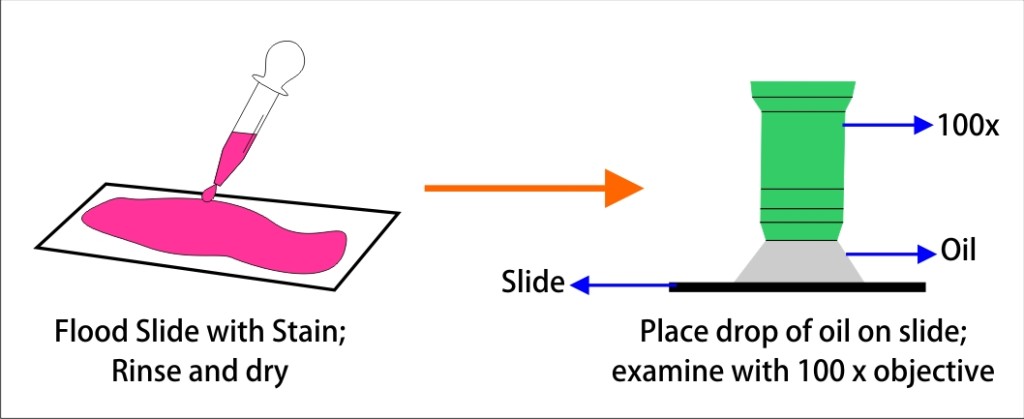
The basic staining process involves fixing or mounting of the sample on to the slide, immersing the sample in dye solution for a specified time period and then finally rinsing and observing the sample under the microscope. In some staining procedures, an additional chemical called mordent is added to the stain which increases the interaction between the cell and the dye forming an insoluble coloured precipitate, which is retained even after the dye is washed away from the cell.
There are basically two types of staining procedures, simple and differential.
Simple staining is the most basic staining performed to determine cell shape, size, and arrangement of bacterial cells. This staining technique stains the bacterial cell. It can be performed with basic dyes with different exposure times. Negative staining is also a simple staining technique, that provides the simplest and probably the quickest technique to get information about the cell shape, cell breakage, and refractile inclusions in cells such as sulphur and poly β hydroxyl butyrate granules and endospores. The bacteria rendered colourless against a coloured background in negative staining. The dyes used in the negative staining are usually anionic in nature and hence repelled by negatively charged cytoplasm, leaving the cells unstained.
Differential Staining is a broad term that is used to describe staining processes which use more than one chemical stain. Differential staining entails use of a series of dyes to stain the different cell organelles based on the structural and compositional differences. This type of staining helps identification and categorization of the cells into different groups. Various differential staining techinques used in microbial studies are
- Gram staining
- Acid fast staining
- Endospore staining
- Capsule staining
- Flagella staining
- Cellwall staining
- Nuclear material staining
Points to Remember:
- A stain is a chemical that adheres to structures of the microorganism and in effect dyes the microorganism so the microorganism can be easily seen under a microscope.
- Basic stains as methylene blue, crystal violet, safranin and malachite green. are cationic, have positive charge and stain chromosomes and the cell membranes of many bacteria.
- Acid stains as eosin and picric acid are anionic, have a negative charge and stain cytoplasmic material and organelles or inclusions.
- A differential stain consists of two or more dyes and is used in the procedure to identify bacteria.
- Most commonly used differential stains for bacterial identification are the Gram stain, the Ziehl-Nielsen acid-fast stain and endospore stain


Responses