Observing Microorganism
Part 1 – Microscopy
Microbiology deals with the study of microorganisms that cannot be seen distinctly with the unaided eye. Observation of microorganisms is an integral part of Microbiology. Considering the nature of the objects to be studied, the microscope becomes an instrument of paramount importance.
Microorganisms are observed and studied with the help of microscopes. The unit of measurement used to measure microorganisms is the Metric System. The size of the specimen determines which microscopes can be used to view the specimen effectively. Modern microscopes produce images with great clarity, magnifications that range from ten to thousands of times.
Types of Microscopes
Simple Microscopes
Simple microscopes have only one lens like a magnifying glass. It has a double convex lens with a short focal length. The examples of this kind of instrument include the hand lens and reading lens.
When an object is kept near the lens, its principal focus with an image is produced, which is erect and bigger than the original object. Leeuwenhoeck’s simple microscopes allowed him to magnify images from 100 to 300 X.
Compound Light Microscopy
These are the most basic type of microscopes used in microbiology. It consists of a series of lenses that utilizes visible light as its source of illumination. Various small specimens can be studied to find details with a compound light microscope.
In a compound light microscope, light originates from an illuminator and passes through condenser lenses, which direct light onto the specimen. The light then enters the objective lenses, which further magnifies the image.

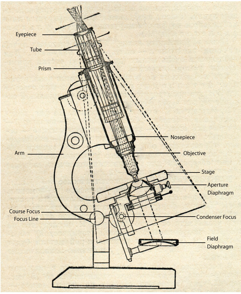
Components of a Compound Microscope
The major components of a compound microscope are :
Framework: The basic frame structure is made up of metal, which includes the arm and base to which whole of the magnification and optical components are attached. The metallic arm is connected to a U shaped strong and heavy base that provides stability to the instrument.
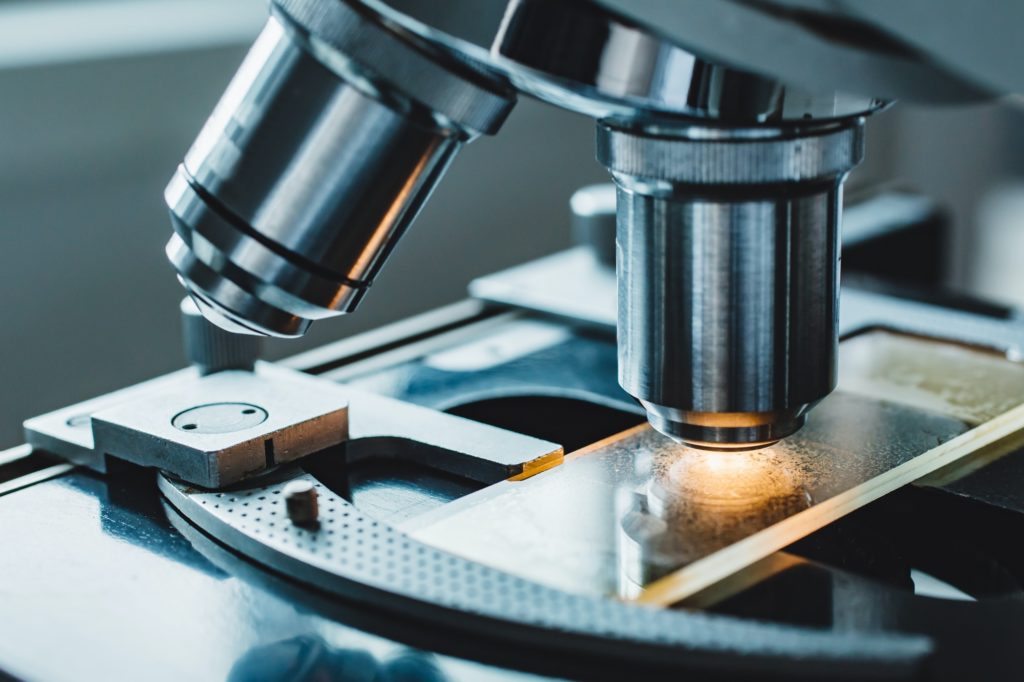
Stage: this is the flat horizontal platform positioned at about halfway through the length of the microscope with a hole at the centre that allows the passage of light for illumination of the sample.
Focus knobs: Two pairs of knobs are attached to the arm that help in up and down movement of the stage and in adjustment and focusing of specimens of different thickness.
Lens Systems: All microscopes employ a set of different types of lens systems: the oculars, the objectives, and the condenser, that have different focusing power, and contribute to the complete magnification system.
Nose piece: A revolving nosepiece which holds the objectives is attached to the curved upper part of the arm of the microscope. The nosepiece can be rotated to position the objective with the required magnification in path of the magnification system, beneath the body assembly and the eye piece.
Eyepiece (ocular lens): The eyepiece or ocular lens is a set of lenses held in a cylindrical tube kept inserted in a tubular structure on the curved upper part of the arm, above the nose piece. It consists of two or more lenses which focus the image into the eye. The newest microscopes consist of a pair of eye pieces that allows the observer to use both the eyes to observe the specimen in the microscope. Such microscopes are called binocular microscopes. The normally used eye pieces have 2X, 50X and 10X magnifications.
Objective: The objectives are usually small cylindrical objects containing a single or a set of lenses attached to the nosepiece. The nosepiece holds three to five objectives, which contain lenses of varying magnifying power (2X-400 X). The total arrangement of the lenses is parfocal, which means that the sample stays in focus even when the lenses are changed from one to another in a microscope.
Condenser: A condenser is also a lens which is fixed below the stage and it focuses the beam of light coming from the light source onto the slide. The condenser is usually aided with diaphragm and/or filters, to control and manage the quality and intensity of the light passing through the sample.
Light Source: The light source is mounted at the base of the microscope. The source of light may be the day light, a halogen light, or even LEDs and lasers, as used in the latest microscopes. The microscopes have some provision for reducing light intensity with a neutral density filter.
Types of Compound Microscopes
- The Bright-Field Microscope – It is the simplest of all the optical microscopy illumination. It helps to see the dark objects against a bright background.
- Dark Field Microscope – This is used to examine live or unstained microorganisms and other specimens like light-sensitive organisms or specimens that lack contrast with their background.
- Phase-Contrast Microscope – It is useful to examine live specimens and does not require fixing or staining, as it can kill or discomfort the living microorganism and will make the observation inaccurate.
- The Differential Interference Contrast Microscope – This type of microscopy takes advantage of differences in the light refraction by different parts of living cells and transparent specimens and allows them to become visible for microscopic evaluation.
- The Fluorescence Microscope – This microscope uses UV light to magnify Fluorescent substances. They can absorb UV light and emit visible light. Sometimes cells are also stained with fluorescent chemicals (fluorochromes) to be studied under this microscope.
- Confocal Microscope – Confocal microscope is majorly used to study the detailed structure of specific objects within the cells.
- Two-Photon Microscope – Also known as two-photon laser scanning microscopy, it is a further refinement of precision fluorescence microscopy.
- Electron Microscopes – It uses electrons, electromagnetic lenses, and fluorescent screens. Electron wavelengths are 100,000 x smaller than visible light wavelength which helps to magnify the specimen. Here the specimens are stained with heavy metal salts to be observed .
All these types of microscopes yield a distinctive image and are used for different types of observation of microorganisms.
How Can We Observe Microorganisms?
The microbial cells are routinely stained for clear microscopic examination. Staining is thus an auxiliary technique used in microscopy and microbiology to enhance contrast in the microscopic images of the microorganisms. Different types of stains and staining procedures are available today to study the multiple properties of various microorganisms.
Stains used in different staining procedures are the aqueous or alcoholic solution of chemical substances known as dyes, which may be natural or synthetic. These stains adhere to the cell and give it colour and contrasts, making the cell more visible. The stains may be acidic, basic or neutral.
The acidic dyes as picric acid, acid fuschin, eosin etc., stain the cytoplasmic components of the cells which are basic in nature, while the basic dyes as methylene blue, crystal violet, safranin etc., stain the acidic components of the cell as nucleic acids. As the bacterial cell is slightly negatively charged on its surface at neutral pH (pH 7), the basic dyes stain bacterial cells as a whole.
The basic staining process involves fixing or mounting of the sample onto the slide, immersing the sample in dye solution for a specified time period and then finally rinsing and observing the sample under the microscope.
In some staining procedures, an additional chemical called mordent is added to the stain which increases the interaction between the cell and the dye forming an insoluble coloured precipitate, which is retained even after the dye is washed away from the cell. There are basically two types of staining procedures, simple and differential.
Simple Staining
Simple staining is the most basic staining performed to determine cell shape, size, and arrangement of bacterial cells. This staining technique stains the bacterial cell. It can be performed with basic dyes with different exposure times.
Negative staining is also a simple staining technique that provides the simplest and probably the quickest technique to get information about the cell shape, cell breakage, and refractile inclusions in cells such as sulphur and poly β hydroxyl butyrate granules and endospores.
The bacteria rendered colourless against a coloured background in negative staining. The dyes used in the negative staining are usually anionic in nature and hence repelled by negatively charged cytoplasm, leaving the cells unstained.
Differential Staining
Differential staining is a broad term that is used to describe staining processes which use more than one chemical stain. Differential staining entails the use of a series of dyes to stain the different cell organelles based on the structural and compositional differences.
This type of staining helps identification and categorization of the cells into different groups. Various differential staining techniques used in microbial studies are
- Gram Staining – Gram staining is a common technique used to differentiate two large groups of bacteria based on their different cell wall constituents. This procedure distinguishes between Gram-positive and Gram-negative groups by colouring the cells either red or violet.
- Acid-fast Staining – It is a differential stain used to identify acid-fast organisms. Acid-fast organisms are characterized by nearly impermeable cell walls as they contain mycolic acid and large amounts of fatty acids, waxes, and complex lipids. As the cell wall is so resistant to most compounds, acid-fast microorganisms require a special staining technique.
- Endospore Staining – It is a differential stain used to visualize bacterial endospores, which are very essential for bacteria as they help to survive in any hostile condition.
- Capsule Staining – This type of staining uses acidic and basic dyes to stain background & bacterial cells so that presence of a capsule is easily visualized.
- Flagella Staining – These stains are mainly prepared to coat the surface of the flagella with dye or a metal such as silver.
- Cell wall Staining – Bacterial cell walls are smeared in the water on a slide and, as soon as the air dries out, it is stained 3-4 minutes with a 1 % aqueous solution of new fuchsin.
- Nuclear Material Staining – Nuclear staining refers to the staining of cell nuclei only. The nucleus contains a large amount of nucleic acid, so a reagent that binds to nucleic acids is used in this type of staining.
Points to Remember:
- Microorganisms are too minute in size to be seen by unaided eyes, hence are observed and studied using microscopes.
- Compound microscopes are commonly used in research labs and institutes to study microorganisms. They use glass lenses to bend and focus light rays and produce enlarged images of small objects.
- Microscopes are delicate and very expensive instruments in any academic or research institutes. Even the most basic compound microscopes require a lot of investment. They, hence, need critical care in usage and handling.
- A stain is a chemical that adheres to structures of the microorganism and in effect dyes the microorganism so the microorganism can be easily seen under a microscope.
- Basic stains as methylene blue, crystal violet, safranin and malachite green are cationic, have a positive charge and stain chromosomes and the cell membranes of many bacteria.
- Acid stains as eosin and picric acid are anionic, have a negative charge and stain cytoplasmic material and organelles or inclusions.
- A differential stain consists of two or more dyes and is used in the procedure to identify bacteria.
- Most commonly used differential stains for bacterial identification are the Gram stain, the Ziehl-Nielsen acid-fast stain and endospore stain.


Responses